Myocardial infarction

Myocardial

Heart disease is one of the leading causes of death in the world. In Thailand the number of deaths from heart disease is increasing each year. The main condition that patients are suffering from is coronary artery disease resulting in myocardial infarction.

Myocardial infarction is the result of the lack of blood supply to the cardiac muscle through atherosclerotic coronary artery disease obstructing the blood flow. The symptoms of myocardial infarction include a crushing pain around the chest, which may spread to the arms, the neck, the jaw, and the molars. Patients who are experiencing a major infarction may have palpitations, feel tired, sweat and lose consciousness and can even die suddenly.

If you have chest pains or suffer from the symptoms mentioned above or feel pain around the chest for 15-20 minutes, see a doctor as soon as possible. Early intervention can save your life.

The doctor will inquire about the patient’s general health, and order medical and blood checks, including EKG. The patient may need an exercise stress test in order to ascertain the fitness of the heart muscle or and echo cardiogram to check the efficiency of the heart. If the doctor finds any abnormal symptoms then a 128-Slice CT Scan may be in order to check coronary arteries. The accuracy of this diagnostic procedure is 90%. These checks are non-invasive, so are painless.

Further treatment depends on the condition of the heart muscle and the reasons for any blockages. Medical treatment with vasodilators may be enough, though interventional techniques including balloon angioplasty or stenting may be necessary or even coronary artery bypass grafts.

The best treatment for cardiac conditions is “prevention” A healthy lifestyle, a healthy diet and an annual check-up if you are above 40 years of age will go a long way towards ensuring that you too do not die from a myocardial infarction.

‘Finally, do not ignore symptoms of heart disease. Early treatment can save your life!’

Out Patient Department (OPD)

Out Patient Department of Bangkok Pattaya Heart Center is dedicated in carrying the highest standard of professional medical care and services to patients suffering from all types of heart disease treated by experienced interventional cardiologists and cardiovascular thoracic surgeons. Moreover, all patients are treated by highly qualified nurses and technicians and the most advanced equipment and technology accessible.

The specialist Heart Center is one of the best of its kind in its field providing the quality and care as expected international standard. The Heart center also receives many patient referrals from numerous other hospitals in the region.

The Heart Center is devoted in delivering every aspect of cardiovascular care, including all of following interventional cardiology, electrophysiology, advanced heart failure and cardiovascular surgery. Of course as is widely known and accepted prevention is better than cure that is the reason why The Heart center uses comprehensive holistic approach ranging from prevention and ensure detection to diagnosis, treatment and rehabilitation.

The Hospital Heart center provides the latest technology and the very best in medical care and service ensuring that you will be guaranteed of the best treatment available and enabling you re-recuperate.

The following facilities are offered:
  • Cardiac Diagnostic Investigation Centre very best
  • Cardiac Imaging ( 64 Slices Spiral CT Scan)
  • Outpatient consultation center
  • Non-Invasive cardiac testing
  • Cardiac catheterization Laboratories
  • Electrophysiology Laboratory
  • Cardiac surgery operating theatres
  • 29 beds dedicated to Coronary Care Unit (CCU)
  • Cardiac Telemetry Monitoring Unit
  • Moblie CCU
  • MRI
Non-Invasive Procedures:
  • Electrocardiogram (EKG) is a test that records the electrical activity of the heart used to measure the rate and regularity of heartbeats
  • Exercise Stress Test (EST) is a general screening tool to test the effect of exercise on heart.
  • Echocardiography (Echo) is an ultrasound of the heart.
  • Transesophageal echocardiography (TEE) is a technique used when an echocardiography does not give enough detailed information.
  • Dobutamine Stress Echocardiography involves taking a medication called dobutamine while you are closely monitored and the medication stimulates your heart and makes it “think” it is exercising.
  • Tilt Table Test (TTT) is designed to use for patients experiencing a malfunction in the central nervous system, which controls the heart.
  • Ankle Brachial Index (ABI) is a simple non-invasive vascular screening test to assess arteriosclerosis by measuring the blood pressure on the arms and legs.
  • Holter Monitoring (DCG) is continuous monitoring of the electrical activity of a patient’s heart muscle ( electrocardiography ) for 24 hours, using a special portable device called a Holter monitor.
Cardiac Care Unit (CCU)

The Bangkok Pattaya Heart Center provide the present 29 bedded coronary care unit adjacent to the cath lab and the cardiac surgical theatre provides all the most advanced life support facilities necessary to salvage a patient with cardiac dysfunction secondary to a heart attack. Basic facilities like thrombolytic therapy is provided with the best available medicines like Urokinase, Streptokinase and Recombinant tissue plamsinogen activator. Facilities of primary angioplasty, defibrillation, pacing, noninvasive and invasive ventilation, intra aortic balloon pump and dialysis and surgical support if needed enhances the chances of survival when one is faced with this life threatening disease.

Cardio Wards

The Bangkok Pattaya Heart Center offer both semi-private and private rooms to suit the patient’s needs. All are tastefully decorated with facilities to ensure comfort and convenience for the patients stay. The advanced equipments such as Telemetry (t he electronic transmission of data from a patient’s heart to a monitoring station) are provided in every room. Well-trained physicians and nursing staffs are on duty 24 hours a day to provide continuous care.

Catheterization Unit (Cath Lab)

The state of the art digital cath lab provides the ideal safe environment for the interventional procedures like PTCA, stenting and balloon valvuloplasty. Many of the cardiac procedures, like atrial septal defect and patent ductus arteriosus which required open-heart surgery earlier, can be safely tackled with interventional procedures at present. The new cath lab provides twice more clarity and minimizes the radiation dose by half thereby enhancing patient safety by four times. The facility is optimal for pace maker insertion as well.

Invasive Procedures:
  • Cardiac Catheterization is a test to check the heart and coronary arteries which is used to check blood flow in the coronary arteries, blood flow and blood pressure in the chambers of the heart.
  • Percutaneous Transluminal Coronary Angioplasty (PTCA & Stent) is to open up peripheral arteries that are narrowed or blocked by plaque build-up (atherosclerosis) or the stenotic vessels by placing a “balloon” with/without a stent into the stenotic area of the blood vessels.
  • Pacemaker is a medical device designed to regulate the beating of the heart which stimulate the heart when either the heart’s native pacemaker is not fast enough or if there are blocks in the heart’s electrical conduction system preventing the propagation of electrical impulses from the native pacemaker to the lower chambers of the heart, known as the ventricles.
  • Automatic Implantable Cardioverter Defibrillator (AICD) is an electronic device like a large pacemaker that is implanted surgically in a pocket formed in the chest wall. It consists of a pulse generator that can deliver a powerful shock to the heart; electrodes to sense the rhythm of the heart and to deliver the shock to the heart muscle; and a computer and circuitry that tells the AICD when to discharge the shock.

Arrhythmia

arrhythmia
What is an arrhythmia?

An arrhythmia is an irregular heart rhythm resulting from an abnormality in the heart’s electrical system. The heart’s electrical system is responsible for setting the pace of your heartbeat. A rhythm that is too fast is called a tachycardia, a rhythm that is too slow is called a bradycardia.

What causes an arrhythmia?

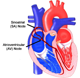
Each contraction of the heart is triggered by electrical impulses sent from the sinoatrial node (SA node) in the right atrium, an upper chamber of the heart. The impulses travel through the atria to the atrioventricular node (AV node), where they are transmitted to the heart’s lower chambers or ventricles.

Arrhythmias may occur due to:
  • An abnormal functioning of the cells in the SA node.
  • Delays in the conduction or receiving of impulses
Heart-related conditions that may lead to arrhythmias include:
  • Damage from a heart attack or atheroselerosis
  • Heart defects or congenital heart disease
  • Abnormal heart structures or functions, such as those resulting from cardiomyopathy or valvular heart disease.
  • Effects from medications
  • Other causes including stress, caffeine or alcohol consumption, smoking, reaction to non-prescription medications or a lack of sleep.
What are the different types of arrhythmias?

There are many types of arrhythmias.arrhythmia

  • Sick sinus syndrome is a condition in which the SA node does not fire often enough, causing the heart rate to slow down.
    There may be alternating periods of rapid heartbeats.
  • Sinus arrhythmias are irregular heart rhythms that also begin in the SA node.
  • Sinus tachycardia is a rapid heart rate caused by electrical impulses firing faster than normal.
  • Heart blockage results when electrical signals to the ventricles are delayed or blocked.
  • Premature ventricular contractions occur when an early signal from a ventricle causes a premature heartbeat. This is often felt as a “skipped beat.”
  • Ventricular fibrillation results from very fast and uncontrollable electrical signals. They cause the heart to quiver rather than beat.
  • Walff-Parkinson-White syndrome occurs when there is an extra connection, or pathway, between the atria and ventricles.
  • Atrial fibrillation is the result of rapid and disorganized signals from the atria that prompt the ventricles to contract irregularly.
  • Atrial flutter is a specific type of atrial tachycardia in which the atria contract regularly, but extremely rapidly.
  • Paroxysmal atril tachycardia is characterized by rapid heartbeats originating in the atria.
  • Premature atrial contractions are premature signals in the atrium that cause the heart to beat too soon.
How are arrhythmias treated?

Treatment of an arrhythmia depends on the type of arrhythmia and its severity. Non sustained arrhythmias may only require certain lifestyle changes, such as:

  • Reducing caffeine intake
  • Limiting the amount of alcohol consumption
  • Quitting smoking
  • Avoiding certain medications, such as some decongestants
  • Using stress management techniques

Medications that may be prescribed include beta-blockers, calcium channel blocke digoxin or antiarrhy-thmics. These medications should be monitored carefully to prevent side effects, which may include increased or more severe arrhythmias. Patients using these medications are encouraged to learn how to take their own pulse in order to detect irregular rhythms.

More invasive procedures may sometimes be necessary, including:
  • An artificial pacemaker for bradycardias
  • An implantable cardioverter defibrillator for abnormally fast heart rhythms.
  • Catheter ablation to destroy abnormal areas of the heart where arrhythmias originate

PERIPHERAL STENTS

Why is the doctor performing this procedure?

To open up a peripheral artery that is narrowed or blocked by plaque build-up (atherosclerosis), and to structurally support that opening by permanently placing a wire stent within the artery.

What is the procedure?

A stent is mesh-like wire cylinder. Stent placement is often part of a PTA (angioplasty) procedure. As in angioplasty, a catheter is inserted into an artery – usually in the groin – but sometimes in the arm or wrist. The catheter is advanced to the blocked peripheral artery, and a series of x-ray pictures are taken to clearly visualize the artery that is narrowed. Then a balloon-tipped catheter is advanced into the narrowed artery. Inside the artery, the balloon is inflated and deflated several times, compressing the plaque against the artery wall and widening the artery so blood flow improves.

This balloon-tipped catheter is removed, and a separate balloon-tipped catheter, with a stent attached, is advanced to the area that was just opened. The balloon is inflated, expanding the stent into the inner layer of the artery. The balloon is removed, but the stent stays in place, acting as a scaffold to keep the artery open. The inner lining of the artery will then heal around the stent.

X-ray pictures are repeated, and if the stent has been successfully placed, the catheters are removed. Pressure is applied to the puncture site (to stop bleeding) while the patient rests quietly.

Where is the procedure performed?

In the Catheterization Lab or Radiology.

How long does this procedure take?

Angioplasty with stent placement usually takes 1-2 hours.

Percutaneous Transluminal Coronary Angioplasty (PTCA)

Why is the doctor performing this procedure?

To open up peripheral arteries that are narrowed or blocked by plaque build-up (atherosclerosis). The peripheral arteries most commonly affected by atherosclerosis are:

  • Cerebral arteries in the brain
  • Carotid arteries in the neck
  • Renal arteries leading to the kidneys
  • Abdominal arteries
  • Iliac arteries in the groin
  • Femoropopliteal arteries in the thigh
  • Infrapopliteal arteries in the knee
What is the procedure?

Percutaneous transluminal angioplasty is commonly called PTA, or just angioplasty. A catheter is inserted into an artery – usually in the groin – but sometimes in the arm or wrist. The catheter is advanced to the blocked artery, and a series of x-ray pictures are taken to clearly visualize the artery that is narrowed. Then a balloon-tipped catheter is advanced into the narrowed artery. Inside the artery, the balloon is inflated and deflated several times, compressing the plaque against the artery wall and widening the artery so blood flow improves.

X-rays pictures are repeated, and if the artery has been successfully re-opened, the catheters are removed. Pressure is applied to the puncture site (to stop bleeding) while the patient rests quietly.

Where is the procedure performed?

In the Catheterization Lab.

How long does this procedure take?

PTA (angioplasty) usually takes 1-2 hours.

How long does the patient stay in the hospital?

1 day in CCU and 1-2 day in Cardio Ward.