Cardiac Catheterization
A Cardiac catherization is a minimally invasive test that offers clear,accurate information about the heart, the coronary arteries located on the surface of the heart and possibly, the aorta. A small tube called a catheter is used to help a physician identify narrowed or clogged arteries, evaluate the heart’s four valves and assess congenital heart defects.
There are four major parts to a cardiac catheterization:
- Measuring blood pressure within the heart’s major arteries
- Taking blood samples for testing
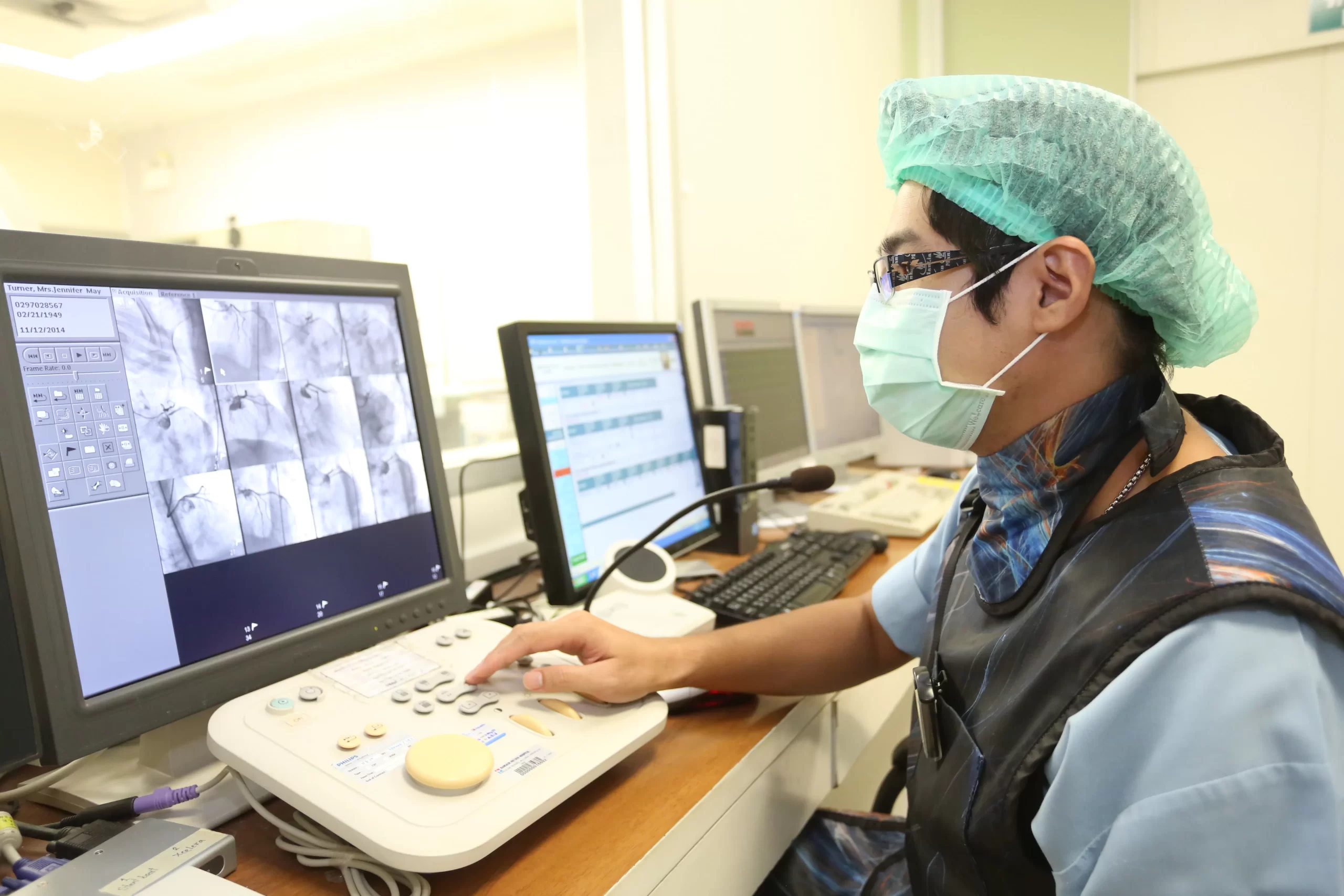
- Performing a coronary angiogram, and x-ray of the coronary arteries
- Performing a left ventriculogram, and x-ray of the lower left chamber of the heart
A cardiac catheterization may be performed for two reasons. First, it may provide important information about the heart and major arteries, help the physician to more clearly see the source of a heart-related problem and determine whether the patient is good candidate for surgery.
In addition, cardiac catheterization are performed to help make a diagnosis. They are usually done after other, less invasive, tests have been performed on patients who may have a heart-related condition. A cardiac catheterization would follow test such as:
- An electrocardiogram (EKG), recording the heart’s electrical activity on a moving strip of paper
- An echocardiogram, using sound waves to create an image of the heart’s structure and function
- An exercise stress test, using EKG technology while the patient exercises on a treadmill or statonary bicycle
How do you prepare for a cardiac catheterization?
Typically, patients will be advised to continue taking most medications as normal, with some exceptions such as anticoagulants and antiplatelets. These medications interfere with the blood’s ability to clot, so dosage may be reduced or the medication suspended at some point prior to the test. Patients are encouraged to discuss their full medication schedule, including over-the-counter drugs and dietary supplements, with their physician so all necessary adjustments can be prescribed.
All patients should supply the physician with copies of previous test results, such as EKGs, if available. Diabetic patients may require further preparation instructions and should seek further guidance from their physician.
On the morning of the test, a light breakfast is suggested. Immediately before the test, patients will be asked various questions about their medical history. Patients should discuss any history they may have regarding blood clotting disorders or allergy reactions to:
- Iodine
- Shellfish
- Strawberries
- Dyes used in previous tests or procedures
Cardiac catheterization may be performed as either an inpatient or outpatient procedure. It takes place in a cool, sterile catheterization laboratory.
The patient is made comfortable, and then an intravenous (I.V.) tube is inserted to deliver a sedative and any other necessary medications. Heart rate and rhythms are continually monitored.
The catheter is usually invented into the femoral artery in the thigh/groin, but some physicians may choose to use the elbow or wrist. It is then fed toward the heart, which could cause some minor discomfort.
When the catheter reaches the target area, blood pressure measurements and, possibly, blood samples will be taken. A dye is injected through the catheter. This injection could cause a brief feeling of nausea, a headache, palpitations or a flushing/reddening across the body.
The physician will usually perform the ventriculogram first, followed by a coronary angiogram and, in some cases, an aortogram.
The test typically takes 30 minutes to one hour.
Share :