Arthroscopy is a treatment that is used to diagnose and treat a variety of knee problems. A surgeon makes a small incision about the size of a buttonhole and inserts a tiny camera. A surgeon can discover abnormalities inside your knee by viewing the inside your joint on a high-definition video monitor.
Knee arthroscopy is a frequent minimally invasive surgical treatment. Using pencil-thin surgical devices inserted through additional small incisions, surgeons can even heal some types of joint injury during arthroscopy.
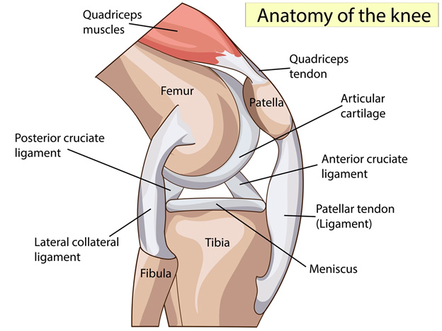
Anatomy
Your knee is your body’s largest and most intricate joint. The lower end of the femur (thighbone), the upper end of the tibia (shinbone), and the patella (kneecap) are the bones that make up the knee.
The following are some of the other essential structures that make up the knee joint:
- Articular cartilage. Articular cartilage covers the extremities of the femur and tibia, as well as the back of the patella. As you bend or straighten your leg, this slick substance helps your knee bones move smoothly across one another.
- Synovium. Synovium is a thin lining that surrounds the knee joint. This lining secretes a lubricant that coats the cartilage and decreases friction during movement.
- Meniscus. Shock absorbers are two wedge-shaped sections of meniscal cartilage located between the femur and tibia. The meniscus, unlike articular cartilage, is strong and springy and serves to cushion and stabilize the joint.
- Ligaments. Ligaments connect the bones to one another. The four main ligaments in your knee work together like strong ropes to keep the bones in place and keep your knee stable.
- Your knee has two collateral ligaments, one on each side.
- The two cruciate ligaments are located within the knee joint. The anterior cruciate ligament in front and the posterior cruciate ligament in rear cross each other to form an X.
Anatomy of the knee in its natural state. Arthroscopy is a procedure that is used to diagnose and treat abnormalities with the articular cartilage, ligaments, and other structures that surround the joint.
Who needs knee arthroscopy?
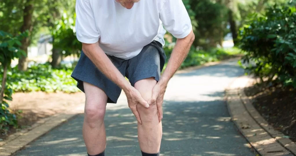
If you’re experiencing knee discomfort that isn’t improving with non surgical therapy, your doctor may consider a knee arthroscopy. Nonsurgical alternatives include rest, ice, nonsteroidal anti-inflammatory drugs, and physical therapy (PT). Despite the fact that osteoarthritis is caused by arthritis, arthroscopic knee surgery isn’t always a successful treatment.
Arthroscopic surgery can be used to diagnose and treat a variety of knee ailments, including the following:
- torn anterior or posterior cruciate ligaments
- meniscus tear (the cartilage between the bones in the knee)
- patella that’s out of position
- loose fragments of torn cartilage in the joint
- removal of a Baker’s cyst
- fractures in the knee bones
- synovium swollen (the lining in the joint)
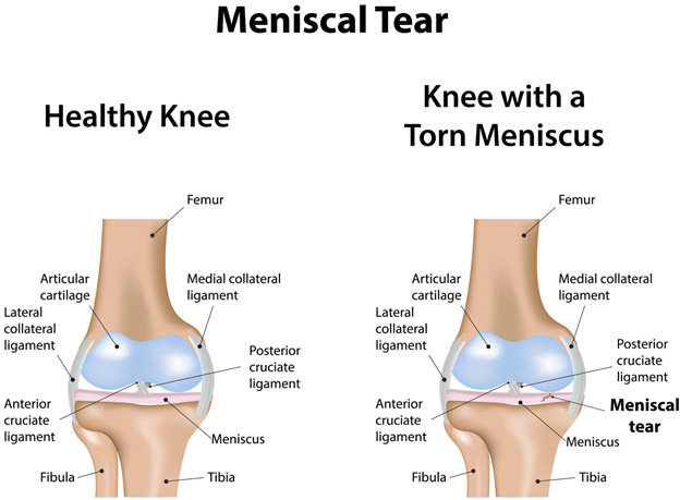
(Left) A Healthy Knee
(Right) A flap tear is a significant meniscal tear.
Surgery
Evaluations and Tests
Before your operation, your orthopedic surgeon may ask that you see your primary care physician to check your overall health. They’ll decide if there are any issues that could prevent the process from going well. A more thorough examination may be required before surgery if you have certain health risks.Preoperative testing may be ordered by your orthopedic surgeon to aid in the planning of your treatment. Blood tests or an electrocardiogram are two examples (EKG).
Admissions Instructions
Your knee arthroscopy will most likely be done as an outpatient treatment if you are otherwise healthy. This implies you won’t have to stay in the hospital overnight.
Make sure your orthopedic surgeon is aware of any medications or supplements you are taking. Some of these medications may need to be stopped prior to surgery.
The hospital will call you ahead of time to discuss your operation in greater detail. Follow the directions for when to arrive and, more importantly, when to cease eating and drinking before your surgery.
Anesthesia
A member of the anesthesia team will speak with you prior to your procedure. Local, regional, or general anesthesia can be used during knee arthroscopy:
- Local anesthesia numbs just your knee
- Regional anesthesia numbs the lower body.
- General anesthesia puts you to sleep
Which procedure is ideal for you will be discussed with you by your orthopedic surgeon and anesthesiologist.
Surgical Procedure
The surgeon will create a few small incisions in your knee, known as portals, to begin the operation. To fill the knee joint and rinse away any murky fluid, a sterile solution will be utilized. This allows your orthopedic surgeon to see the internal structures of your knee in amazing detail.
The first duty for your surgeon is to correctly diagnose your condition. The arthroscope will be inserted and guided by the image presented on the screen. Your surgeon will put tiny instruments through other small incisions if surgery is required.
Shaving, cutting, grabbing, and meniscal healing all require specialized devices. Stitches are commonly anchored to the bone using special devices.
The arthroscope and surgical equipment will be inserted through small incisions called portals by your surgeon.
Complications
Following arthroscopic surgery, the chances of complications are relatively low. If issues do arise, they are usually minimal and simple to cure. The following are some of the possible side effects of knee arthroscopy:
- Infection
- Blood clots
- Knee stiffness
- Accumulation of blood in the knee
- Bruising or swelling
Recovery
You will be transported to the recovery room after surgery and should be able to go home after 1 or 2 nights. Make arrangements for someone to drive you home and check on you the first evening.
While a knee arthroscopy recovery is quicker than a standard open knee surgery recovery, it is critical to carefully follow your doctor’s recommendations after you get home.
Pain Management
You will have some discomfort following surgery. This is a common occurrence during the healing process. Your doctor and nurses will manage to help you lessen your pain, allowing you to recover more quickly from surgery.
Following surgery, medications are frequently provided for short-term pain management. There are several medications available to assist control pain, your doctor may also prescribe a combination of drugs.
Everyone has a different reaction to surgery. Inquire with your healthcare practitioner when you will be able to resume your normal activities, such as driving and walking without assistance. Before engaging in more physically demanding activities, your doctor may urge you to wait a few weeks.
People’s lifestyles and levels of activity must sometimes be altered. Knee injuries can occur during certain activities, notably those that demand you to sprint or leap. Ask your doctor about low-impact sports and activities that are easier on your knees.
Risk and Benefit
Knee arthroscopy advantages
Traditional (open) surgery usually takes longer to recuperate from than less invasive procedures like knee arthroscopy. You’ll be able to get back on your feet faster than with standard surgery because only a few minor stitches are required. You may also have less pain and a lower risk of infection.
What are the risks or complications of knee arthroscopy?
Complications from knee arthroscopy are rare, however there are risks specific to a knee arthroscopy, such as:
- bleeding inside the knee joint
- formation of a blood clot in the leg
- infection inside the joint
- stiffness in the knee
- injury or damage to the cartilage, ligaments, meniscus, blood vessels, or nerves of the knee