Digital Mammogram with Tomosynthesis

breast center04

breast center04

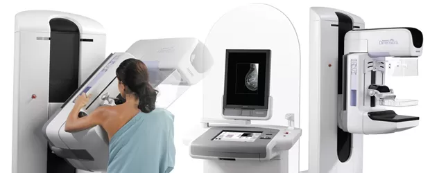
A specific type of breast imaging that can produce results in 2D and 3D at the same time, taking about 10 seconds for each image and get a good result that helps detect cancer and makes diagnosis between the breast lump and tumor more accurate. Radiation oncologist can read the image in each dimension or each slices out a reconstruction image to analyze the shape and size of suspected lump. It can diagnose the difference between fat, connective tissue and lymphatic system to find small sized calcium that can signal the presence of cancer. Staging of the condition is a significant factor in the treatment as ‘the earlier the condition is found the more chance of cancer recover and treatment’.

3D Digital Mammogram comes with a low compression unlike the old device (X-ray) as the image goes in +/- 15º and displays a result in 3D. This makes the procedure less painful. Not only the radiation oncologist can read the result more accurate but also reduce recall rates and improves the quality of patients identification for breast biopsy of each patients.

Women age from 35 years old are advised to have breast examination every year or according to the specialist’s recommendation.


Advantages of 3 Tesla MRI in Musculoskeletal Imaging

mri001

Clinical musculoskeletal MR imaging is now transitioning from 1.5T to 3T systems.

The advantages of higher field strength in the evaluation of musculoskeletal disorders are apparent since the better signal-to-noise ratio (SNR) and contrast-to-noise ratio (CNR) can be applied in multiple fashions to improve imaging times and image resolution.

The potential for demonstrating and enhancing the visibility of several osseous, tendinous, cartilaginous, ligamentous and other intrinsic joint structures in faster scan time has been appreciated. Such clearer visualization of small structures and the pathology leads to potentially increased diagnostic accuracy. The potential of coil technology and parallel imaging for 3T MRI also helps produce superb images comparing to 1.5T systems. Excellent image quality has impressed referring orthopedic surgeons and rheumatologists, plus the reduction in scan time has resulted in greater patient satisfaction and reduced anxiety. It has been shown that 3T MR imaging vastly improves diagnostic abilities in the musculoskeletal system, which means better patient care and management.

Better Image Quality, High Resolution

Better Positioning, More Comfort, 70 cm bore

Better Technology, Digital Broadband MR