Test for bacteria in the stomach (C-13 Urea Breath Test)

C13UBT EN

C13UBT EN

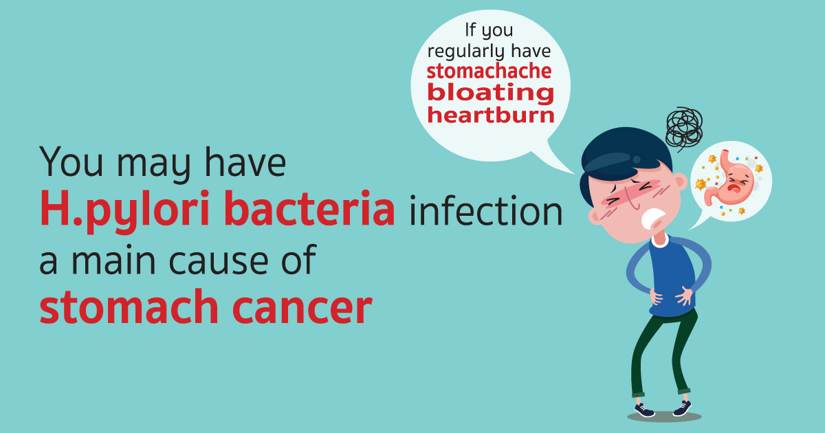
Helicobacter pylori bacteria (H. pylori) in the stomach is an important cause of pathology in the stomach and upper colon (duodenum). In addition, H. pylori bacteria is a risk factor for gastric cancer. The test can be done by blowing into a device that looks like a small bag without the need of endoscopy and takes about 20 minutes and the result analysis takes about 30 minutes measuring carbon dioxide (13 Co2) caused by the reaction of H. pylori bacteria that has Urease enzyme into blood stream then expel to the lungs and outside by the breath using the principle of IR Spectrophotometer which is 95-100% accurate

Currently, there are many ways to test for H. pylori which are:

  • Urea test from biopsy obtained from Gastroscopy
  • Histology examination which requires special straining
  • Culture and serology examination which is not currently popular

At present, there is no report of side effects or danger from the test as it is relatively safe method. It can test in children and pregnant women. If you have frequent stomachache and do not test for the bacteria you may be at risk of gastritis, gastric ulcer, duodenal ulcer, gastric cancer in the future.

H. pylori bacterial infection in the stomach is still a major problem in the Thai population. Lifestyle behavior, food consumption, living in crowded environment community area or living with those who have bacteria infection in the stomach but do not show symptoms all is at great risk. H. pylori bacteria cause disease and complications that are dangerous to the body in many ways. When the H. pylori bacteria is detected, you should be treated with antibiotics and repeat test to confirm that you are completely free from infection. This is to prevent the recurrence of gastric ulcer including reduction of the risk of gastric cancer as well.

What is FibroScan?

fibro12

What is Fibroscan?
It is a newly ultrasound based technology that visualizes tissue and fats in the liver. The examination is painless, and reduces any chance of infection compared to ‘Liver Biopsy

How does Fibroscan work?
Vibration Controlled Transient Elastography (VCTE) uses low frequency waves at 50 hertz into to calculate any fibrous tissue in your liver.
Controlled Attenuation parameter (CAP) is used to calculate fats in the liver.

Symptoms and possible causes of Liver problems

  • Jaundice and Fatigue
  • Digestion Dysfunction
  • Chronic Alcoholism
  • Family members with Liver Cancer
  • Hepatitis B and C

Does it hurt?

  • No!
  • The entire procedure is over within 5-10 minutes’
  • Slight vibration when the probe is on the skin surface.
  • No problems with serial examinations.

Having Fibroscan

  • Lie down with both arms are above your head. The technician applies gel on the probe or patient skin.
  • The examination takes 10 readings to get the final result.
  • The doctor will take the result and advise any treatment required.

What is Fibroscan for?
It helps in diagnosis and evaluation to plan the treatment. It is a better option for the patients who do not wish for a liver biopsy.

Who can carry out Fibroscan procedures?
Only Gastroenterologists and Nuclear medicine specialists can process the test.

Cautions with Fibroscan

  • It only can be used for Liver diagnosis
  • Cardiac patient who has a pacemaker and defibrillator
  • Patient with ascites (fluid in the abdomen
  • Not recommended during pregnancy

For more information, please contact GI and Liver Center Tel. 0 3890 9345

Investigations

Gastroscopy

Gastroscopyis a medical term that has two parts: gastro for “stomach” and scopy for “looking”. Gastroscopy is a diagnostic test that enables the doctor to view the stomach. The instrument used to perform this simple test is the gastroscope: a long, thin, flexible fiberoptic tube. Within the end of this remarkable device is a miniaturized color TV camera with a wide angle lens. By passing this “scope” through the stomach, your doctor can directly examine the lining of your upper digestive system. The examination is quick, painless and without incision.

Colonoscopy

Colonoscopy allows the physician to investigate the entire large intestine, from the lowest part, to the rectum, all the way up through the colon to the lower end of the small intestine. This procedure is used to detect early signs of cancer in the colon and rectum. It can also be used to diagnose the causes of changes in bowel habits. Colonoscopy enables the physician to detect inflamed tissue, abnormal growths, ulcers and bleeding

Virtual colonoscopy (VC)

Virtual colonoscopy is a medical imaging procedure which uses x-rays and computers to produce two – and tree-dimensional images of the colon (large intestine) from the lowest part, the rectum, all the way to the lower end of the small intestine and display them on a screen. Virtual Colonoscopy provides clearer, more detailed images than a conventional x-ray using a barium enema, sometime called a lower gastrointestinal (GI) series. It also takes less time than either a conventional colonoscopy or a lower GI series. Since it does not use a colonoscopy, no sedation is needed and the patient can return to his/her usual activities or go home after the procedure without the aid of another person.

CP (Endoscopic Retrograde Cholangiopancreatography)

Diagnostic ERCP (endoscopic Retrograde Cholangiopancreatography):

Identifies a problem within the bile duct or pancreas e.g. gallstones, cancer of the bile ducts, chronological pancreatitis.

Early Detection of GI-Cancer

The newest innovation for early detection of GI cancer is called “Narrow Band Imaging (NBI)”, an optical filter technology that radically improves the visibility of capillaries, veins and other subtle tissue structures, by optimizing the absorbance and scattering characteristics of light. This method is used in the upper and lower digestive system details up to the cellular level, requires no NBI images on the monitor also help doctors to indicate areas of abnormality without waiting for pathology analysis.

Gastrointestinal Wireless Capsule Endoscopy is a new medical technological advancement for diagnosis of small intestinal neoplasms in patients with gastrointestinal polyposis syndrome. The examination is quick, painless, without incision, small risk, with very high accuracy. It was found to be one of the most convenient, safe, and effective methods for detecting small intestine problems. This technology has already been trialled in many countries including USA, UK Germany, Italy, Israel, and Hong Kong.

Common diseases can easily occur in the Gastro-Intestinal system.