Hybride operatiekamer

Hybrid OR

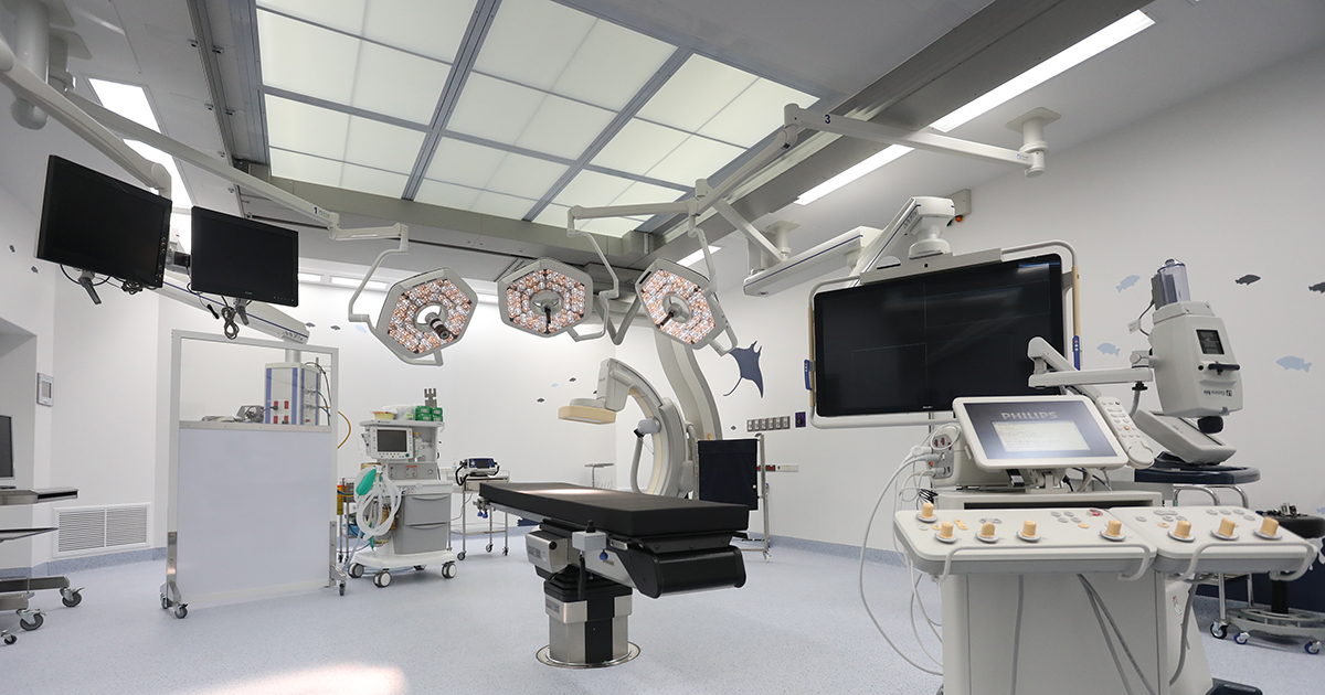
Een stap vooruit in de medische technologie voor patiënten met vaatziekten waarvoor chirurgie de primaire behandelingsoptie is. Door tijdens de operatie gebruik te maken van hoogwaardige radiologische techniek, geïnstalleerd in de operatiekamer, is er sprake van verdergaande mogelijkheden bij gecompliceerde chirurgische behandelingen. De kenmerken van een hybride operatiekamer zijn:

  • Ontworpen met extra ruimte om met een multidisciplinair team (zoals een chirurg, radioloog, cardioloog, anaesthesist, radiologische technici, verpleegkundigen) systematisch te kunnen werken
  • De laatste state-of-the-art operatie gereedschappen en een vasculair röntgenapparaat met een helder beeld om nauwkeurig de plaats, omvang en pathologie van het vasculaire probleem vast te stellen. De operatietafel is ontworpen om de straling uit alle invalshoeken bij de patiënt te laten doordringen.
  • Uitgerust met een set van anaesthetische inhalatietoestellen, medisch gas verdelers en een aan het plafond bevestigd medisch uitrustingsrek met een chirurgische lamp voorzien van een gelode spiegel voor goed zicht tussen de controlekamer en de operatiekamer
  • Een luchtcirculatiesysteem dat de verspreiding van stofdeeltjes in de lucht beperkt en daarmee het infectierisico reduceert. Uiteraard heeft de operatiekamer stralingsbestendige muren en is de vloer voorzien van een materiaal dat micro-organismen afstoot hetgeen de veiligheid van patiënt en personeel in de operatiekamer verhoogd.

Voordelen van het ondergaan van een procedure in de hybride operatiekamer

  1. Diagnose en behandeling in één bezoek spaart tijd. Kortere opname, minder bloedverlies, voorspoediger herstel
  2. Geschikt voor patiënten met complexe aandoeningen met grote risico’s. De multidisciplinaire aanpak reduceert deze risico’s aanzienlijk
  3. Noodzakelijke ingrepen met een kleine chirurgische wond, ingrepen met een vasculaire katheter door de huid zoals dotteren te zamen met algemene chirugie

Het gebied van deze vorm van chirurgie

  • Orgaantransplantatie
  • Cardiovasculaire en thoracale operaties
  • Diagnose en chirurgie voor aandoeningen gerelateerd aan het vasculaire systeem
  • Diagnose en laparoscopische chirurgie bij gynaecologische en urologische aandoeningen
  • Diagnose, chirurgie en procedures met hoogwaardig technologische hulpmiddelen door een gespecialiseerd team

Mechanische Trombectomie

MechanicalThrombectomy1

MechanicalThrombectomy1

Behandeling van een ischemisch of acuut herseninfarct door middel van mechanische trombectomie.

Mechanische trombectomie is tegenwoordig een zeer effectieve behandeling voor de verwijdering van bloedproppen die de doorstroming van bloed in de aderen in de hersenen blokkeren. Zodoende worden de bloedvaten weer geopend en dus uw hersenen weer van bloed voorzien. Deze behandelingsmethode is niet alleen erg effectief maar reduceert ook het risico op invaliditeit als gevolg van zuurstoftekort in de hersenen of hersenoedeem zoals bij een klassieke craniotomie.

Met deze methode kan het ondergaan van hersenchirurgie worden voorkomen en zijn ook de kansen op hersenkneuzing, invalideit en zelfs overlijden voor patiënten met trombose in de hersenen kleiner.

De indicaties voor patiënten die voor mechanische trombectomie in aanmerking komen, zijn:

  • Patiënten die een behandeling hebben ondergaan met intraveneuze fibrinolyse medicijnen, die geen verbetering hebben ondergaan
  • Patiënten met een bloedprop in de hersenslagader
  • Patiënten die geen fibrinolyse medicijnen kunnen krijgen of abnormale bloedklontering hebben

Patiënten met tekenen van stenose of verstopping zoals duizeligheid, zwakte, gedeeltelijke verlamming, problemen met het bewegen van de tong, wazig zicht, verlies van balans moeten onmiddellijk naar het ziekenhuis.

Volgens het zogenaamde “Stroke Fast Track system” kan een patiënt met een herseninfarct binnen 24 uur worden geholpen met mechanische trombectomie door een team van neurologen en vasculaire chirurgen in een gereedstaande hybride operatiekamer.

Thrombectomie Mécanique

MechanicalThrombectomy1

MechanicalThrombectomy1

Traitement de l’AVC ischémique ou aigu par thrombectomie mécanique

La thrombectomie mécanique est un traitement très efficace aujourd’hui, qui élimine les caillots sanguins qui bloquent l’artère cérébrale et libère les vaisseaux sanguins qui permettent au sang de nourrir le cerveau.

Ce traitement sera plus efficace pour le patient atteint de caillots dans les artères cérébrales. Il réduit également les incapacités dues aux œdèmes cérébraux et cérébraux ischémiques pendant la craniotomie.

Cette méthode aidera à réduire les risques pour le patient de subir une intervention chirurgicale, une commotion cérébrale, une invalidité et le décès chez les patients atteints de thrombose des artères cérébrales.

Voici les indications pour les patients nécessitant une thrombectomie mécanique

  • Les patients qui ont été traités avec des médicaments de fibrinolyse par voie intraveineuse, sans amélioration.
  • Patients présentant un caillot sanguin dans la grande artère cérébrale.
  • Les patients incapables de prendre des médicaments fibrinolytiques ou présentant une coagulation sanguine anormale
  • Les patients présentant des signes de risque de sténose ou d’occlusion, d’étourdissements, de faiblesse, de demi-hémisphère ou des deux côtés, de difficultés à bouger la langue, de vision floue, de perte d’équilibre, doivent être immédiatement conduits à l’hôpital.

Soins d’urgence «Le système Stroke Fast Track» convient au traitement des patients ayant subi un AVC avec thrombectomie mécanique avec la collaboration de neurologues et de chirurgiens vasculaires, en utilisant une salle d’opération hybride adaptée à tous les traitements accélérés et prête à effectuer une thrombectomie mécanique en moins de 6 heures.

What is FibroScan?

fibro12

What is Fibroscan?
It is a newly ultrasound based technology that visualizes tissue and fats in the liver. The examination is painless, and reduces any chance of infection compared to ‘Liver Biopsy

How does Fibroscan work?
Vibration Controlled Transient Elastography (VCTE) uses low frequency waves at 50 hertz into to calculate any fibrous tissue in your liver.
Controlled Attenuation parameter (CAP) is used to calculate fats in the liver.

Symptoms and possible causes of Liver problems

  • Jaundice and Fatigue
  • Digestion Dysfunction
  • Chronic Alcoholism
  • Family members with Liver Cancer
  • Hepatitis B and C

Does it hurt?

  • No!
  • The entire procedure is over within 5-10 minutes’
  • Slight vibration when the probe is on the skin surface.
  • No problems with serial examinations.

Having Fibroscan

  • Lie down with both arms are above your head. The technician applies gel on the probe or patient skin.
  • The examination takes 10 readings to get the final result.
  • The doctor will take the result and advise any treatment required.

What is Fibroscan for?
It helps in diagnosis and evaluation to plan the treatment. It is a better option for the patients who do not wish for a liver biopsy.

Who can carry out Fibroscan procedures?
Only Gastroenterologists and Nuclear medicine specialists can process the test.

Cautions with Fibroscan

  • It only can be used for Liver diagnosis
  • Cardiac patient who has a pacemaker and defibrillator
  • Patient with ascites (fluid in the abdomen
  • Not recommended during pregnancy

For more information, please contact GI and Liver Center Tel. 0 3890 9345

Investigations

Gastroscopy

Gastroscopyis a medical term that has two parts: gastro for “stomach” and scopy for “looking”. Gastroscopy is a diagnostic test that enables the doctor to view the stomach. The instrument used to perform this simple test is the gastroscope: a long, thin, flexible fiberoptic tube. Within the end of this remarkable device is a miniaturized color TV camera with a wide angle lens. By passing this “scope” through the stomach, your doctor can directly examine the lining of your upper digestive system. The examination is quick, painless and without incision.

Colonoscopy

Colonoscopy allows the physician to investigate the entire large intestine, from the lowest part, to the rectum, all the way up through the colon to the lower end of the small intestine. This procedure is used to detect early signs of cancer in the colon and rectum. It can also be used to diagnose the causes of changes in bowel habits. Colonoscopy enables the physician to detect inflamed tissue, abnormal growths, ulcers and bleeding

Virtual colonoscopy (VC)

Virtual colonoscopy is a medical imaging procedure which uses x-rays and computers to produce two – and tree-dimensional images of the colon (large intestine) from the lowest part, the rectum, all the way to the lower end of the small intestine and display them on a screen. Virtual Colonoscopy provides clearer, more detailed images than a conventional x-ray using a barium enema, sometime called a lower gastrointestinal (GI) series. It also takes less time than either a conventional colonoscopy or a lower GI series. Since it does not use a colonoscopy, no sedation is needed and the patient can return to his/her usual activities or go home after the procedure without the aid of another person.

CP (Endoscopic Retrograde Cholangiopancreatography)

Diagnostic ERCP (endoscopic Retrograde Cholangiopancreatography):

Identifies a problem within the bile duct or pancreas e.g. gallstones, cancer of the bile ducts, chronological pancreatitis.

Early Detection of GI-Cancer

The newest innovation for early detection of GI cancer is called “Narrow Band Imaging (NBI)”, an optical filter technology that radically improves the visibility of capillaries, veins and other subtle tissue structures, by optimizing the absorbance and scattering characteristics of light. This method is used in the upper and lower digestive system details up to the cellular level, requires no NBI images on the monitor also help doctors to indicate areas of abnormality without waiting for pathology analysis.

Gastrointestinal Wireless Capsule Endoscopy is a new medical technological advancement for diagnosis of small intestinal neoplasms in patients with gastrointestinal polyposis syndrome. The examination is quick, painless, without incision, small risk, with very high accuracy. It was found to be one of the most convenient, safe, and effective methods for detecting small intestine problems. This technology has already been trialled in many countries including USA, UK Germany, Italy, Israel, and Hong Kong.

Common diseases can easily occur in the Gastro-Intestinal system.

Digital Mammogram with Tomosynthesis

breast center04

breast center04

A specific type of breast imaging that can produce results in 2D and 3D at the same time, taking about 10 seconds for each image and get a good result that helps detect cancer and makes diagnosis between the breast lump and tumor more accurate. Radiation oncologist can read the image in each dimension or each slices out a reconstruction image to analyze the shape and size of suspected lump. It can diagnose the difference between fat, connective tissue and lymphatic system to find small sized calcium that can signal the presence of cancer. Staging of the condition is a significant factor in the treatment as ‘the earlier the condition is found the more chance of cancer recover and treatment’.

3D Digital Mammogram comes with a low compression unlike the old device (X-ray) as the image goes in +/- 15º and displays a result in 3D. This makes the procedure less painful. Not only the radiation oncologist can read the result more accurate but also reduce recall rates and improves the quality of patients identification for breast biopsy of each patients.

Women age from 35 years old are advised to have breast examination every year or according to the specialist’s recommendation.


Advantages of 3 Tesla MRI in Musculoskeletal Imaging

mri001

Clinical musculoskeletal MR imaging is now transitioning from 1.5T to 3T systems.

The advantages of higher field strength in the evaluation of musculoskeletal disorders are apparent since the better signal-to-noise ratio (SNR) and contrast-to-noise ratio (CNR) can be applied in multiple fashions to improve imaging times and image resolution.

The potential for demonstrating and enhancing the visibility of several osseous, tendinous, cartilaginous, ligamentous and other intrinsic joint structures in faster scan time has been appreciated. Such clearer visualization of small structures and the pathology leads to potentially increased diagnostic accuracy. The potential of coil technology and parallel imaging for 3T MRI also helps produce superb images comparing to 1.5T systems. Excellent image quality has impressed referring orthopedic surgeons and rheumatologists, plus the reduction in scan time has resulted in greater patient satisfaction and reduced anxiety. It has been shown that 3T MR imaging vastly improves diagnostic abilities in the musculoskeletal system, which means better patient care and management.

Better Image Quality, High Resolution

Better Positioning, More Comfort, 70 cm bore

Better Technology, Digital Broadband MR