Cardiac Catheterization

A Cardiac catherization is a minimally invasive test that offers clear,accurate information about the heart, the coronary arteries located on the surface of the heart and possibly, the aorta. A small tube called a catheter is used to help a physician identify narrowed or clogged arteries, evaluate the heart’s four valves and assess congenital heart defects.

There are four major parts to a cardiac catheterization:

  • Measuring blood pressure within the heart’s major arteries
  • Taking blood samples for testing
  • Performing a coronary angiogram, and x-ray of the coronary arteries
  • Performing a left ventriculogram, and x-ray of the lower left chamber of the heart

A cardiac catheterization may be performed for two reasons. First, it may provide important information about the heart and major arteries, help the physician to more clearly see the source of a heart-related problem and determine whether the patient is good candidate for surgery.

In addition, cardiac catheterization are performed to help make a diagnosis. They are usually done after other, less invasive, tests have been performed on patients who may have a heart-related condition. A cardiac catheterization would follow test such as:

  • An electrocardiogram (EKG), recording the heart’s electrical activity on a moving strip of paper
  • An echocardiogram, using sound waves to create an image of the heart’s structure and function
  • An exercise stress test, using EKG technology while the patient exercises on a treadmill or statonary bicycle
How do you prepare for a cardiac catheterization?

Typically, patients will be advised to continue taking most medications as normal, with some exceptions such as anticoagulants and antiplatelets. These medications interfere with the blood’s ability to clot, so dosage may be reduced or the medication suspended at some point prior to the test. Patients are encouraged to discuss their full medication schedule, including over-the-counter drugs and dietary supplements, with their physician so all necessary adjustments can be prescribed.

All patients should supply the physician with copies of previous test results, such as EKGs, if available. Diabetic patients may require further preparation instructions and should seek further guidance from their physician.

On the morning of the test, a light breakfast is suggested. Immediately before the test, patients will be asked various questions about their medical history. Patients should discuss any history they may have regarding blood clotting disorders or allergy reactions to:

  • Iodine
  • Shellfish
  • Strawberries
  • Dyes used in previous tests or procedures

Cardiac catheterization may be performed as either an inpatient or outpatient procedure. It takes place in a cool, sterile catheterization laboratory.

The patient is made comfortable, and then an intravenous (I.V.) tube is inserted to deliver a sedative and any other necessary medications. Heart rate and rhythms are continually monitored.

The catheter is usually invented into the femoral artery in the thigh/groin, but some physicians may choose to use the elbow or wrist. It is then fed toward the heart, which could cause some minor discomfort.

When the catheter reaches the target area, blood pressure measurements and, possibly, blood samples will be taken. A dye is injected through the catheter. This injection could cause a brief feeling of nausea, a headache, palpitations or a flushing/reddening across the body.

The physician will usually perform the ventriculogram first, followed by a coronary angiogram and, in some cases, an aortogram.

The test typically takes 30 minutes to one hour.

What is FibroScan?

fibro12

What is Fibroscan?
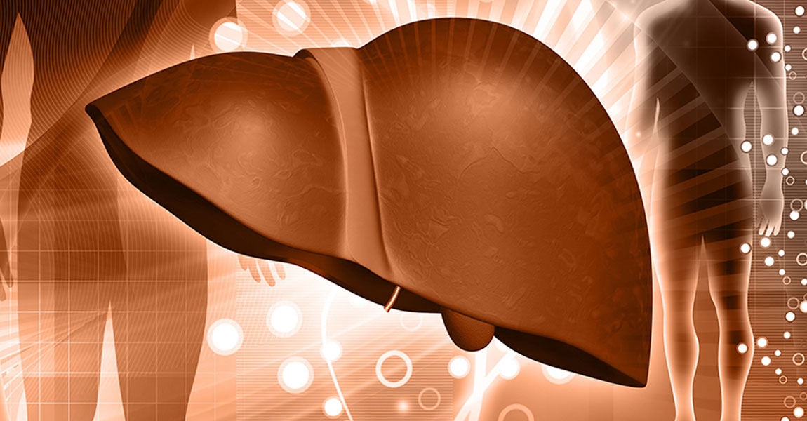
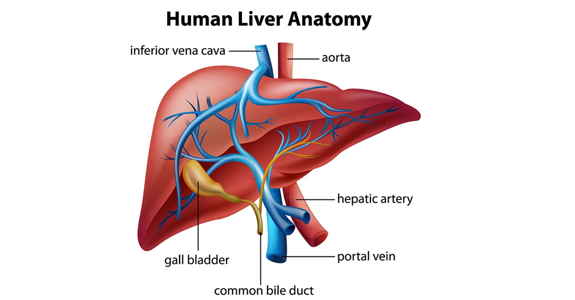
It is a newly ultrasound based technology that visualizes tissue and fats in the liver. The examination is painless, and reduces any chance of infection compared to ‘Liver Biopsy

How does Fibroscan work?
Vibration Controlled Transient Elastography (VCTE) uses low frequency waves at 50 hertz into to calculate any fibrous tissue in your liver.
Controlled Attenuation parameter (CAP) is used to calculate fats in the liver.

Symptoms and possible causes of Liver problems

  • Jaundice and Fatigue
  • Digestion Dysfunction
  • Chronic Alcoholism
  • Family members with Liver Cancer
  • Hepatitis B and C

Does it hurt?

  • No!
  • The entire procedure is over within 5-10 minutes’
  • Slight vibration when the probe is on the skin surface.
  • No problems with serial examinations.

Having Fibroscan

  • Lie down with both arms are above your head. The technician applies gel on the probe or patient skin.
  • The examination takes 10 readings to get the final result.
  • The doctor will take the result and advise any treatment required.

What is Fibroscan for?
It helps in diagnosis and evaluation to plan the treatment. It is a better option for the patients who do not wish for a liver biopsy.

Who can carry out Fibroscan procedures?
Only Gastroenterologists and Nuclear medicine specialists can process the test.

Cautions with Fibroscan

  • It only can be used for Liver diagnosis
  • Cardiac patient who has a pacemaker and defibrillator
  • Patient with ascites (fluid in the abdomen
  • Not recommended during pregnancy

For more information, please contact GI and Liver Center Tel. 0 3890 9345

Investigations

Gastroscopy

Gastroscopyis a medical term that has two parts: gastro for “stomach” and scopy for “looking”. Gastroscopy is a diagnostic test that enables the doctor to view the stomach. The instrument used to perform this simple test is the gastroscope: a long, thin, flexible fiberoptic tube. Within the end of this remarkable device is a miniaturized color TV camera with a wide angle lens. By passing this “scope” through the stomach, your doctor can directly examine the lining of your upper digestive system. The examination is quick, painless and without incision.

Colonoscopy

Colonoscopy allows the physician to investigate the entire large intestine, from the lowest part, to the rectum, all the way up through the colon to the lower end of the small intestine. This procedure is used to detect early signs of cancer in the colon and rectum. It can also be used to diagnose the causes of changes in bowel habits. Colonoscopy enables the physician to detect inflamed tissue, abnormal growths, ulcers and bleeding

Virtual colonoscopy (VC)

Virtual colonoscopy is a medical imaging procedure which uses x-rays and computers to produce two – and tree-dimensional images of the colon (large intestine) from the lowest part, the rectum, all the way to the lower end of the small intestine and display them on a screen. Virtual Colonoscopy provides clearer, more detailed images than a conventional x-ray using a barium enema, sometime called a lower gastrointestinal (GI) series. It also takes less time than either a conventional colonoscopy or a lower GI series. Since it does not use a colonoscopy, no sedation is needed and the patient can return to his/her usual activities or go home after the procedure without the aid of another person.

CP (Endoscopic Retrograde Cholangiopancreatography)

Diagnostic ERCP (endoscopic Retrograde Cholangiopancreatography):

Identifies a problem within the bile duct or pancreas e.g. gallstones, cancer of the bile ducts, chronological pancreatitis.

Early Detection of GI-Cancer

The newest innovation for early detection of GI cancer is called “Narrow Band Imaging (NBI)”, an optical filter technology that radically improves the visibility of capillaries, veins and other subtle tissue structures, by optimizing the absorbance and scattering characteristics of light. This method is used in the upper and lower digestive system details up to the cellular level, requires no NBI images on the monitor also help doctors to indicate areas of abnormality without waiting for pathology analysis.

Gastrointestinal Wireless Capsule Endoscopy is a new medical technological advancement for diagnosis of small intestinal neoplasms in patients with gastrointestinal polyposis syndrome. The examination is quick, painless, without incision, small risk, with very high accuracy. It was found to be one of the most convenient, safe, and effective methods for detecting small intestine problems. This technology has already been trialled in many countries including USA, UK Germany, Italy, Israel, and Hong Kong.

Common diseases can easily occur in the Gastro-Intestinal system.